


Anton van Leeuwenhoek was born on October 24, 1632. To earn a living, he was a merchant, and then a cashier, and a storekeeper. However, what he is best known for is his microscope.
The Leeuwenhoek Microscope
Devices to magnify had been discovered prior to Leeuwenhoek, but Leeuwenhoek’s microscope had unusually high magnifying power. His microscopes used one lens, and yet with that one lens he achieved wonders.
Some people believed that he laboriously ground the lenses into shape with a secret method, however the commonly accepted view indicated that such a feat would have been impossible. Come to find out, advances in imaging allowed researchers to view the microscopes without harming them. They found that there was no special method of lens blowing. Leeuwenhoek was exceptionally gifted at grinding tiny lenses.¹
Leeuwenhoek’s Protozoa
Leeuwenhoek studied all sorts of interesting things, including hair, muscle fibers, nerves, blood, and entire insects; but he gained most of his renown for his discovery and research of protozoa. He sent his protozoa discoveries to the Royal Society of London. However, they refused to believe in the existence of protozoa, until upon Leeuwenhoek’s persistence, they sent a team of doctors, jurists, and a vicar to visit him and see for themselves the results of his research. When the team looked through his microscope, they were amazed at what they saw. The team reported back, and the Society insisted that he work for them.
For fifty years thereafter, Leeuwenhoek made observations with his microscope and sent them to scientific institutions including the Royal Society of London. He continued to research in spite of serious illness, and refused to stop until his death in 1723.
The Modern Microscope

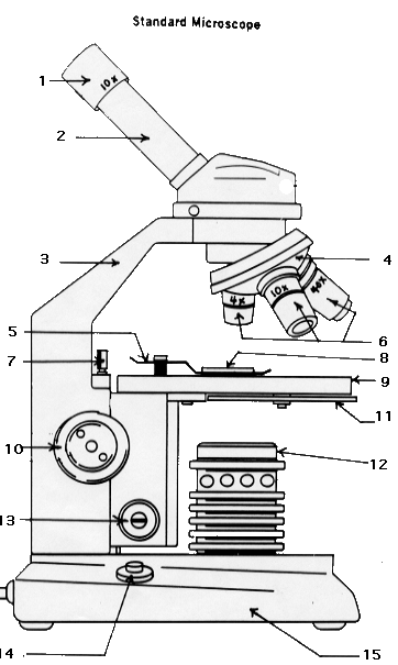
Modern microscopes use multiple lenses to achieve superb magnification. For every lens added, the microscope’s magnification multiplies. For example, if the eyepiece has a magnification of 15 times, and the objective lens (which is the lens that focuses on the object being studied) has a magnification of 40 times, the result is 600 times of amplification (the two numbers being multiplied).
The eyepiece (No. 1 shown in the illustration to the right) usually does have a magnification of around 15 times, while the objective piece (No. 3) varies enormously. Often enough, a bank of objective lenses of different values are attached to a rotating disk (No. 2), allowing the magnification to be changed. A light source (No. 7) under the object illuminates it, providing a sharp image. To focus on an object, adjustments are provided which bring the object being studied closer to or farther from the objective lenses.
Leeuwenhoek’s Microscope Compared to a Modern Microscope
As for Leeuwenhoek’s microscope, it doesn’t look much like anything you see today. A large copper plate held the tiny eyepiece, while a needle was used to hold the specimen before it. The needle was adjusted as needed.
However, to change the magnification, Leeuwenhoek had to build a whole new microscope! The instrument was crude, but highly effective compared to anything else available at the time.
Leeuwenhoek’s Legacy
As can be seen, Leeuwenhoek’s microscope revolutionized the field of microscopes. It proved that large magnifications were possible, and opened the door to detailed study of living things significantly smaller than the period at the end of this sentence.
That study is what we now call microbiology.
¹ Delft University of Technology, “Mystery of superior Leeuwenhoek microscope solved after 350 years,” Phys.org, March 22, 2018, https://phys.org/news/2018-03-mystery-superior-leeuwenhoek-microscope-years.html.
Further Investigation
Anton Van Leeuwenhoek
A biography from PBS.
Tiny Lenses
All about the lenses used in Leeuwenhoek’s microscope.
History of the Microscope
Brief history that also includes a photo of a van Leeuwenhoek microscope.
The Microscope – Parts and Specifications
Diagram with explanations.
Amoeba
Information at EnchantedLearning.com.
Ron’s Pond Scum
A vast series of images taken through a microscope. “An Adventure in Protozoan Art.”
Activities
How to Use a Microscope
Interactive from Wisconsin Technical College includes an exercise on identifying the parts.
Let’s Make an Antoni van Leeuwenhoek’s Microscope
Great idea at FunSci.com … for the brave!
Microscopic Experiments and Labs
Eighteen microscope experiments at GreatScopes.com including one on protozoa.
Amoeba Coloring
Information and labeling diagram worksheet at BiologyCorner.com.
Virtual Electron Microscope
Interesting interactive at MyScope-Explore.org that lets you use an electron microscope to identify samples.
Books

All in a Drop How Antony van Leeuwenhoek Discovered an Invisible World by Lori Alexander
Award winning title and great first biography of Leeuwenhoek.

The World of the Microscope
Usborne Science and Experiments title that includes basic information such as the history of the microscope, types of microscopes and how a microscope works, but also helpful resources such as how to use a microscope, first projects (to become familiar with slide preparation), drawing what you see, making sections, staining, and mounting and measuring. Activities include examining cells, fungi, simple organisms, microscopic water life, plants, insects, rocks and minerals, and crystals. Perfect for a do-it-yourself science course!

How to Know the Protozoa by Theodore Jahn
Definitive classic on the subject with extensive illustrations. Works very well as part of an older student’s biology course.
The Romance of the Microscope {Free eBook}
Public domain title that includes the history of the microscope and what to look at under the microscope.

Greg’s Microscope by Millicent E. Selsam
Simple text but interesting for early reader. Illustrated by Arnold Lobel.
The Microscope by Andrew Ross
Public domain work for the older student, describing in relatively easy-to-understand language how the instrument works.
Common Objects of the Microscope {Free eBook}
Handy tool for those investigating microscopic marvels.
Unit Studies & Lesson Plans
Van Leeuwenhoek’s Surprise
Lesson plan for learning how microbes were first discovered.
Printables & Notebooking Pages
Standard Microscope
Microscope part labeling diagram for notebook at Miami University (answers).
Label Amoeba Diagram
At EnchantedLearning.com.
Microscope Lab Journal
At GreatScopes.com.
Leeuwenhoek’s Microscope Notebooking Pages
Simple pages for copywork, narrations, or wrapping up.


You must be logged in to post a comment.